Echocardiography
Non-invasive cardiac imaging for function, structure, and remodeling
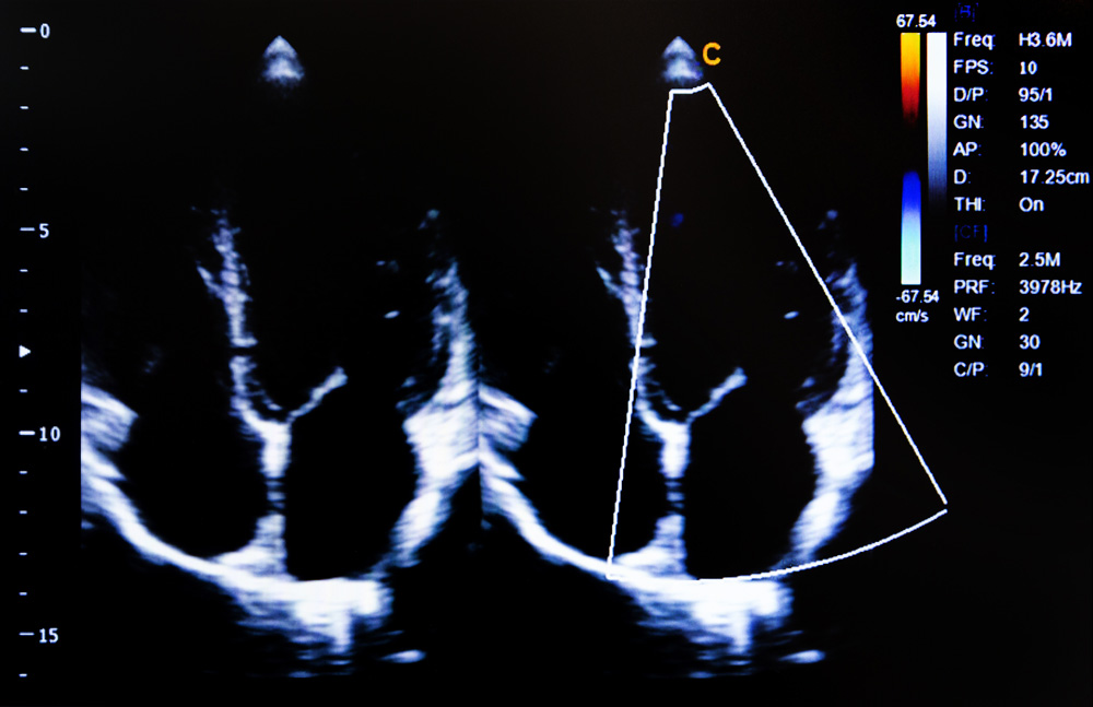
Echocardiography enables high-resolution, real-time visualization of cardiac anatomy and function. CorDynamics integrates echo imaging into both exploratory and IND-enabling studies to support cardiac safety, efficacy, and mechanistic evaluation across disease models.
Preclinical Echocardiography Studies
As a non-invasive modality, echocardiography allows for longitudinal assessment of cardiac function in the same animal over time—minimizing variability and maximizing translational insight. It is routinely used across CorDynamics models, including heart failure, PAH, and ischemia/reperfusion. Capabilities include:
- Cardiac Structure & Function: Ejection fraction, fractional shortening, wall thickness, and chamber dimensions
- Doppler Imaging: Blood flow velocity, diastolic function, and valve performance
- Strain Analysis: Regional myocardial deformation and early detection of subtle dysfunction
- Right Heart Evaluation: RV size, function, and pulmonary pressure surrogates (particularly in PAH models)
Echocardiography is used to:
- Track disease progression and remodeling
- Assess treatment effects in both acute and chronic studies
- Support safety pharmacology and mechanistic discovery
- Complement invasive hemodynamics, telemetry, or electrophysiology endpoints
Why Choose CorDynamics
CorDynamics incorporates echocardiography into a wide range of cardiovascular studies, from discovery-stage efficacy to GLP safety pharmacology. Our experienced team performs high-resolution imaging in both anesthetized and conscious models, using validated imaging protocols and integrated data acquisition systems.
Strain imaging and right heart analysis enhance sensitivity, particularly in subtle models like HFpEF and PAH.
We deliver high-resolution echo imaging to track structure, function, and therapeutic response across various models.


